A coronary calcium CT is usually performed to answer one question:
How much coronary artery calcification does this patient have?
But a CT scan contains considerably more biological information than the single measurement for which it was acquired.
Muscle density, visceral fat, liver attenuation, bone density, lung abnormalities and other features may all be present within the same dataset. The difficulty has traditionally been that extracting these additional measurements manually is impractical.
Artificial intelligence is changing that.
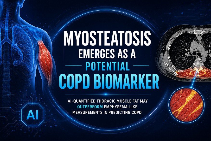
A new study using data from the Multi-Ethnic Study of Atherosclerosis (MESA) suggests that one such opportunistic measurement—thoracic skeletal muscle myosteatosis—may provide information about a patient's future risk of chronic obstructive pulmonary disease (COPD).
The finding is intriguing because the potential biomarker is not located in the lungs.
It is in skeletal muscle.
COPD may be more than a disease of the lungs
COPD is a major global health problem and, according to the World Health Organization, was the third leading cause of death worldwide in 2023, accounting for approximately 3.4 million deaths.
Smoking remains an important risk factor, but COPD is not simply a consequence of smoking exposure. Age, environmental pollutants, impaired physical activity, systemic inflammation, metabolic dysfunction and other factors contribute to disease susceptibility and progression.
This broader perspective raises an interesting question:
Could abnormalities outside the lungs reveal vulnerability to COPD before conventional pulmonary abnormalities become obvious?
The new MESA analysis provides an early indication that the answer may be yes.
What is myosteatosis?
Myosteatosis refers to excessive fat infiltration within skeletal muscle and is generally regarded as a marker of impaired muscle quality.
It differs from simply having a low muscle mass.
Two individuals can have similar muscle volume but very different muscle composition. One may have relatively dense, healthy muscle, whereas the other may have substantial lipid infiltration and therefore lower CT attenuation.
CT can detect this difference because fat has lower attenuation than muscle.
The problem is measurement.
Subtle differences in muscle attenuation are difficult to assess consistently by visual inspection, particularly when thousands of examinations need to be analysed.
This is where AI becomes useful.
Turning an ordinary CT into a quantitative dataset
The study by Azimi and colleagues analysed baseline coronary artery calcium CT examinations from 5,535 participants in MESA.
The cohort had a mean age of approximately 62 years, and 47.6% were male. Importantly, more than half of the participants had never smoked, making the population useful for investigating risk beyond smoking exposure.
The researchers used an AI-based system to automatically quantify thoracic skeletal muscle attenuation.
Myosteatosis was defined using sex-specific attenuation thresholds corresponding to the lowest quartile of thoracic skeletal muscle density:
-
Men: <33.5 HU
-
Women: <27.0 HU
The same CT examinations were also analysed for an emphysema-like lung biomarker, defined by the proportion of lung voxels below −950 HU.
The participants were then followed for approximately two decades.
The outcome was striking.
What did the researchers find?
During the 20-year follow-up period, 396 participants, or 7.1%, were diagnosed with COPD according to hospital discharge records.
Participants with myosteatosis had a substantially greater risk of subsequently receiving a COPD diagnosis.
Before adjustment for other risk factors, the hazard ratio for COPD was approximately 5.98 when the lowest muscle-density quartile was compared with the highest.
After adjustment for age, sex, smoking status, body mass index, race, asthma, physical activity, inflammatory markers and insulin resistance, the association remained significant:
Adjusted HR: 2.74
95% CI: 1.81–4.16
For comparison, the emphysema-like lung measurement had an adjusted hazard ratio of approximately 1.50.
Thus, within this cohort, the muscle-based biomarker demonstrated a stronger statistical association with future COPD than the emphysema-like measurement derived from the same limited CT examination.
That is the central finding.
But it needs to be interpreted carefully.
Why is this finding important?
The most interesting aspect is not that muscle fat is associated with COPD.
It is that a systemic imaging phenotype may contain predictive information that is not obvious from the lungs alone.
Myosteatosis has previously been associated with metabolic dysfunction, frailty and adverse cardiovascular outcomes. COPD, meanwhile, is increasingly understood within the context of systemic inflammation, physical inactivity, metabolic abnormalities and multimorbidity.
These processes may therefore intersect.
A patient with substantial muscle fat infiltration may have evidence of a broader biological phenotype involving impaired metabolic health, inflammation, reduced physical reserve and cardiopulmonary vulnerability.
The CT is simply providing a window into that phenotype.
Why AI matters
Radiologists can recognise severe muscle wasting.
What is much harder is reliably quantifying subtle differences in muscle attenuation across thousands or millions of CT examinations.
AI changes the economics of measurement.
Instead of asking a radiologist to manually identify muscles, place regions of interest and calculate attenuation, an automated system can potentially perform the measurement consistently across an entire imaging database.
This creates something more important than an image.
It creates a quantitative biomarker.
A CT performed for coronary calcium can potentially become a multidimensional health assessment containing measurements of:
coronary calcium + muscle quality + visceral adiposity + liver fat + bone density + lung abnormalities + other imaging biomarkers.
The examination was acquired for one purpose.
AI can extract information relevant to many others.
Why might muscle quality predict lung disease?
The study does not establish the biological mechanism linking myosteatosis to COPD.
Several possibilities are plausible.
Skeletal muscle quality reflects systemic metabolic health. Fat infiltration within muscle can accompany insulin resistance, inflammation and physical inactivity. Reduced muscle quality may also contribute to lower exercise capacity and physical deconditioning.
COPD itself is associated with systemic manifestations extending beyond the lungs, including skeletal muscle dysfunction.
There may therefore be a bidirectional relationship.
Poor muscle quality could be a marker of an underlying systemic environment that predisposes to pulmonary disease, while early respiratory limitation could contribute to reduced physical activity and subsequent deterioration in muscle quality.
The present study cannot determine which process comes first.
That question requires longitudinal studies specifically designed to investigate causality.
An especially interesting finding: smoking was not the whole explanation
One of the more provocative aspects of the analysis was that the association between myosteatosis and future COPD persisted after adjustment for smoking and other established risk factors.
The finding also remained present across subgroups involving age, sex, obesity, smoking history and physical activity.
This does not mean that smoking is unimportant.
Smoking remains a major cause of COPD, and prevention of tobacco exposure remains fundamental. WHO estimates that tobacco smoking accounts for more than 70% of COPD cases in high-income countries.
Rather, the study suggests that imaging-derived muscle quality may capture additional information that conventional risk factors do not completely explain.
That is a more useful interpretation than saying myosteatosis "causes" COPD.
Could this change how radiologists read CT scans?
Potentially—but not yet routinely.
The conventional radiology report is generally focused on the indication for the examination.
For a coronary calcium CT, that means coronary calcification and relevant extracardiac findings.
For a lung CT, it may include pulmonary nodules, emphysema, airway abnormalities and other thoracic findings.
The emerging concept of opportunistic imaging expands this framework.
If an AI system can reliably quantify clinically relevant features without requiring additional imaging, then a single CT examination could become a source of multiple risk markers.
This is already happening in other areas.
AI-based opportunistic assessment of liver steatosis from coronary calcium CT has been investigated for prediction of cardiovascular outcomes and mortality.
Myosteatosis could become another component of this expanding imaging phenotype.
But should every patient with myosteatosis be screened for COPD?
Not on the basis of this study alone.
This is an important distinction.
The study demonstrates prediction, not proof that AI-detected myosteatosis should be used as a population-wide COPD screening test.
There are several reasons.
First, COPD was identified through hospital discharge diagnostic codes rather than systematic spirometric testing of every participant.
Second, the study used a particular CT acquisition and AI-derived measurement.
Third, the optimal threshold for clinical decision-making has not been established.
Fourth, the study was conducted within the MESA cohort and requires external validation in other populations.
Finally, an imaging biomarker is clinically useful only if acting on it improves outcomes.
That last point is often overlooked.
A biomarker can predict disease beautifully and still fail to improve patient care.
What would validation look like?
The next stage is not simply another retrospective analysis.
A stronger validation pathway would involve independent cohorts representing different ethnicities, ages, smoking patterns, body compositions and health-care systems.
Researchers would need to determine:
-
whether myosteatosis predicts spirometric COPD;
-
whether it predicts accelerated decline in FEV1;
-
whether it identifies individuals who later develop respiratory symptoms;
-
whether the association remains after accounting for socioeconomic and environmental exposures;
-
whether serial worsening of myosteatosis corresponds to worsening pulmonary function;
-
and whether incorporating myosteatosis meaningfully improves existing COPD risk models.
The crucial question is ultimately:
Does the addition of myosteatosis change clinical decisions in a way that improves outcomes?
The radiologist's incidental finding may become a risk profile
Imagine a patient undergoing coronary calcium CT.
The scan produces a calcium score.
But the same AI pipeline identifies:
-
moderate coronary calcification;
-
increased visceral adiposity;
-
hepatic steatosis;
-
low thoracic muscle attenuation;
-
reduced bone density;
-
and subtle emphysema-like changes.
None of these findings individually may have been the original reason for the scan.
Together, however, they describe something much more interesting:
the patient's systemic health phenotype.
This is where opportunistic AI could become genuinely transformative.
The future radiology report may not simply describe what is anatomically abnormal.
It may increasingly quantify risk.
From incidental finding to actionable biomarker
There is, however, a danger in producing an ever-growing list of AI-derived measurements.
More biomarkers do not necessarily mean better medicine.
A clinically useful biomarker must be interpretable and actionable.
If an AI system reports that a patient has severe myosteatosis, what should happen next?
Perhaps the appropriate response is assessment of physical activity, nutritional status and metabolic risk.
Perhaps the patient should undergo spirometry if respiratory symptoms or other COPD risk factors are present.
Perhaps cardiovascular risk should be reassessed.
Perhaps the finding should simply be documented and followed.
Clinical pathways must be established before automated measurements become routine.
Otherwise, opportunistic AI could create a new problem: incidentaloma overload at a population scale.
The bigger idea: the CT scan as a digital phenotype
The most important lesson from this research may extend beyond COPD.
Modern CT contains far more information than humans can routinely extract.
Every voxel carries numerical information.
The challenge is converting that information into clinically meaningful measurements.
AI provides the computational layer needed to do this at scale.
The result could be a transition from conventional radiology, where the primary task is lesion detection and characterisation, towards computational phenotyping.
A single CT examination could potentially provide quantitative information about:
atherosclerosis
adiposity
muscle quality
bone health
liver fat
lung structure
vascular calcification
and other systemic markers.
The patient does not need another scan.
The information is already present.
COPD may therefore be hiding in plain sight
The lungs may not always be the first place where future COPD vulnerability becomes measurable.
That is the provocative implication of the MESA study.
Myosteatosis is not yet a validated clinical biomarker for COPD screening, and the findings require external and prospective validation. But the association is strong enough to suggest that muscle composition deserves further investigation as part of the cardiopulmonary risk phenotype.
The larger lesson is perhaps even more important.
Radiology has traditionally been about finding disease in the organ being examined.
Artificial intelligence may allow us to ask a different question:
What else can this scan tell us about the patient?
A coronary calcium CT may contain information about the lungs.
The lungs may contain information about systemic disease.
Muscle may contain information about future cardiopulmonary risk.
And a single examination may ultimately become a quantitative map of health rather than a snapshot of one organ system.
That is the promise of opportunistic imaging.
Not simply seeing more.
Measuring more.
Predicting more.
And, if future research proves the clinical benefit, intervening earlier.